Spatial Sample guidelines
Supported tissues: Here you can find the supported protocols for FFPE and Fresh Frozen (FF) sample preparation. Alternatively, fixed tissue embedded in OCT (Fixed Frozen FXF) can also be processed but is not supported by 10X Genomics. Ensure that your FFPE tissue has been properly fixed, PFA fixation should not exceed 24 hours at 4 oC.
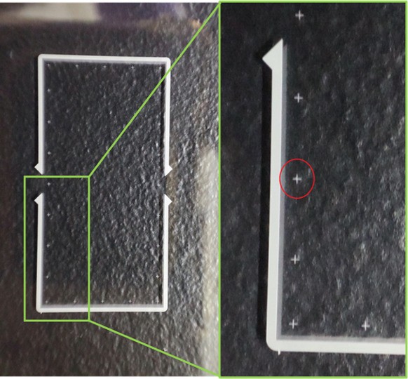
Tissue placement: The capture area of the Xenium slide is 10 x 22 mm. Avoid covering the fiducials with tissue as this risks failures to initiate Xenium Analyzer run. We strongly recommend to practice tissue placement on a blank slide. Print Xenium slide diagram.

Slide storage after tissue placement: After drying, slides containing FFPE tissue can be stored in a desiccator at room temperature for up to 4 weeks. Slides containing FF tissue can be stored at -80°C in a sealed slide mailer for up to 4 weeks.
Tissue slides should be free of wrinkles, folds, air bubbles or cracks.
Quality controls: When performing 5K prime Xenium, the following is required:
1. DV200 test to assess RNA integrity. This should be greater than 30%.
2. On a separate slide, perform an H&E stain and check for tissue artefacts such as: cracks, hemorrhaging and/or improper fixation.
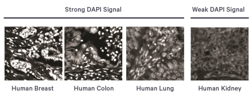
3. On a separate slide, perform a DAPI stain and look for punctate nuclei that are clearly defined.

Supported tissues: Visium HD is a whole transcriptome probe-based gene expression assay compatible with FFPE, Fresh Frozen (FF), & Fixed Frozen tissue (FxF) sections. Here you can find the tested FFPE, FF and FxF and tissues.
Prior to section placement, draw an outline of the allowable area on the back of the blank slide to ensure downstream compatibility. Use slides recommended by 10X
Each Visium HD slide has 2 capture areas of 6.5 x 6.5 mm. When submitting samples to LGTC use a thin marker to annotate your area of interest on the back side of the tissue slide. One tissue slide is required per capture area.
Slide storage after tissue placement: After drying, slides containing FFPE tissue can be stored at 4°C in a desiccator for up to 6 months. Slides containing Fresh Frozen or Fixed frozen (FxF) can be store at -80 oC for up to 2 months.
Imaging guidelines: Follow the H&E or IF imaging guidelines accordingly. When performing IF staining optimization of the antibody concentration is required.
Find here the Visium HD mouse and human probe sets.
Quality controls:
1. Use an adjacent section to measure DV200 and assess RNA integrity. This should be greater than 30%. FFPE and FF RNA extraction kit
2. On a separate slide, perform an H&E stain and check for tissue artefacts such as: cracks, hemorrhaging and/or improper fixation (optional).
3. On a separate slide, perform a DAPI stain and look for punctate nuclei that are clearly defined (optional).
Visium HD 3′ is single cell-scale resolution, unbiased, whole transcriptome, poly-A based gene expression assay compatible with Fresh Frozen tissue sections.
Prior to section placement, draw an outline of the allowable area on the back of the blank slide to ensure downstream compatibility. Use slides recommended by 10X
Slides containing Fresh Frozen can be store at -80 oC for up to 2 months within a slide mailer.
Each Visium HD slide has 2 capture areas of 6.5 x 6.5 mm. When submitting samples to LGTC use a thin marker to annotate your area of interest on the back side of the tissue slide. One tissue slide is required per capture area.
Quality controls:
1. Use an adjacent section (20-30mg tissue) to assess RNA quality by calculating the RIN value using either Bioanalyzer or TapStation. This should be greater than 4. See here recommended RNA isolation kit
2. On a separate slide, perform an H&E stain and check for tissue artefacts such as: cracks, hemorrhaging and/or improper fixation (optional).
3. On a separate slide, perform a DAPI stain and look for punctate nuclei that are clearly defined (optional).
Modifications to documentation (Demonstrated Protocols and User Guide) provided here are unsupported. 10X cannot guarantee assay performance even when implementing the guidance listed in this article and therefore, customers should proceed at their own risk. 10x Genomics Support will not be able to provide additional information beyond this article or replacement reagents.
Tissue sections processed using the Xenium In Situ workflow remain largely intact post-run and can be used for additional applications. Examples of compatible Xenium post-run workflows include immunofluorescence (IF) staining, H&E staining, and whole transcriptome spatial RNA sequencing using the Visium CytAssist Spatial Gene Expression workflow.
Post-Xenium In Situ Applications: Immunofluorescence, H&E, Visium v2, and Visium HD
Post-Xenium Staining: H&E and IF staining samples should be stain within 3 days after the Xenium run.
Because cell segmentation antibodies in the Xenium Multi-Tissue Stain Mix are raised in rabbit, do not use anti-rabbit secondary antibody for post Xenium applications as it will result in cross-detection. For all post-Xenium applications after cell segmentation staining, use primary antibodies that are not raised in rabbits.